Research
The Ostrowski lab integrates several techniques to unravel the underlying mechanisms of autonomic and respiratory dysfunction in Alzheimer’s disease (AD). We are using a non-transgenic rat model to mimic the sporadic form of AD, which affects about 95% of all patients. Small doses of Streptozotocin injected into the lateral ventricles of the rat brain elicits many pathologies commonly observed in human AD, including memory loss, amyloid plaques, and hyperphosphorylated tau. Other commonly observed signs are increased inflammation, oxidative stress, and neuron loss. In addition, the STZ-rat model of AD displays alterations in cardiorespiratory function. Such disturbances may be due to alterations in brainstem nuclei that are important for the maintenance of blood pressure and respiration.
We employ several complementary techniques to unravel the mechanisms behind the alterations seen with AD:
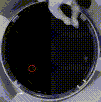
Behavioral tests verify correct induction of our AD model include passive/active/step down avoidance test, plus maze, and Morris water maze. In the latter rats are trained to find a hidden platform in a circular pool using visual cues for orientation. After 24 hours, recall of this learned behavior is considered to be long-term memory.
Plethysmography is used to analyze parameters important for respiration (tidal volume, breath rate, and minute ventilation). We examine the change of these parameters to conditions with reduced oxygen content (hypoxia; e.g. 10% O2) or increased carbon dioxide (hypercapnia; e.g. 5% CO2). Under healthy conditions, hypoxia or hypercapnia upregulate respiration to increase oxygen delivery to the body or eliminating excess CO2 via expiration respectively.
Normoxia (21% O2)
Hypoxia (10% O2)

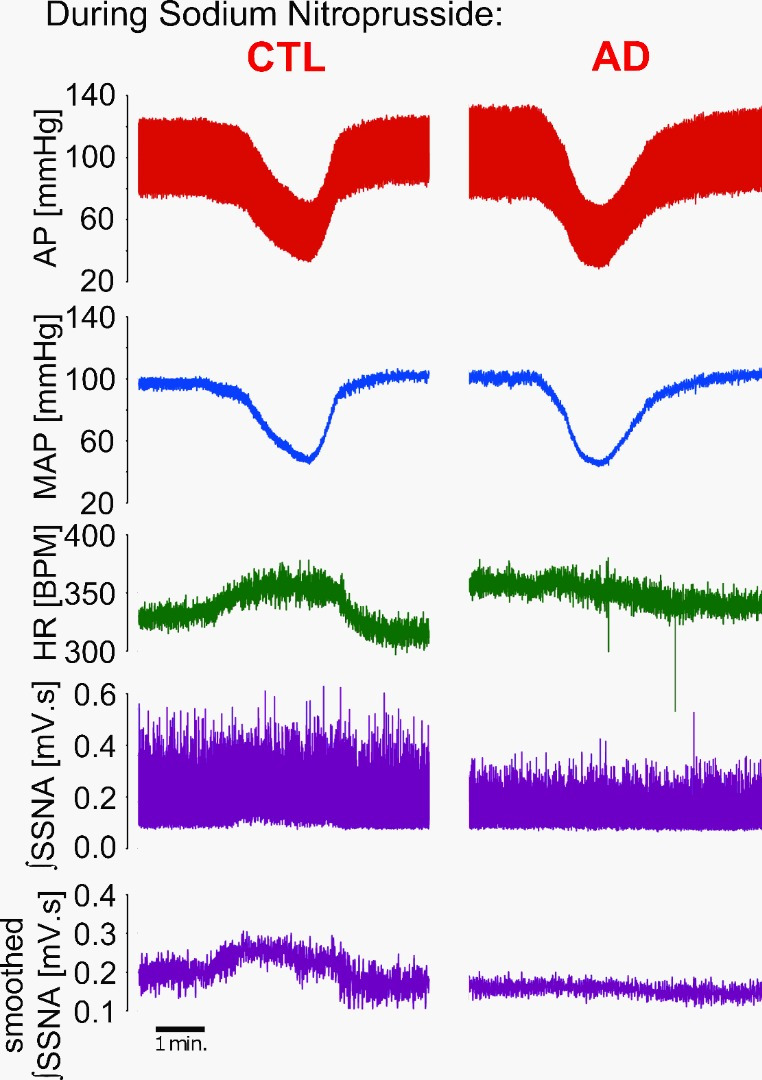
Cardiovascular assessment is done by monitoring responses of the heart or autonomic nervous system to changes in blood pressure. We raise/lower blood pressure by infusion of phenylephrine/sodium nitroprusside via catheters inserted in the femoral vein. Baroreflex-mediated compensatory responses to the change in arterial pressure will then be evident by changes in heart rate and splanchnic sympathetic nerve activity.
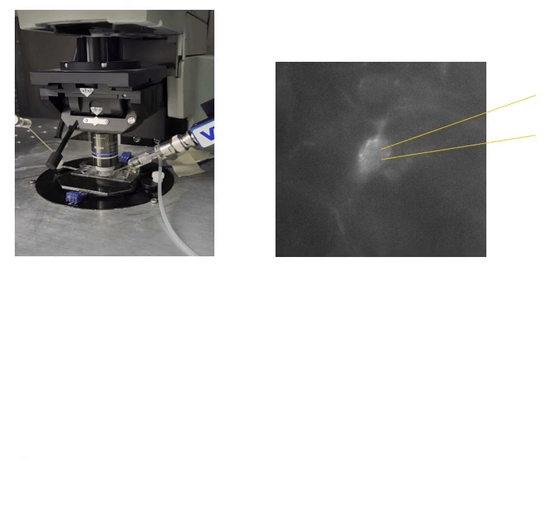
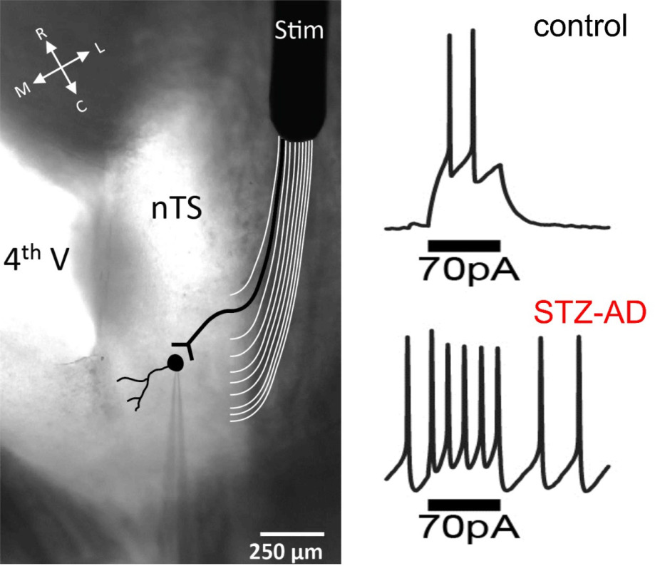
Patch clamp recordings in the brain slice allow us to see neuronal activity in various structures of the brain. A main focus of the Ostrowski laboratory is the nucleus tractus solitarii, a brainstem region that is integral for the control of respiration, blood pressure, and many other physiological functions. Using this technique we are able to analyze network activity of the nucleus, synaptic transmission, as well as the spiking activity and membrane properties of individual neurons.
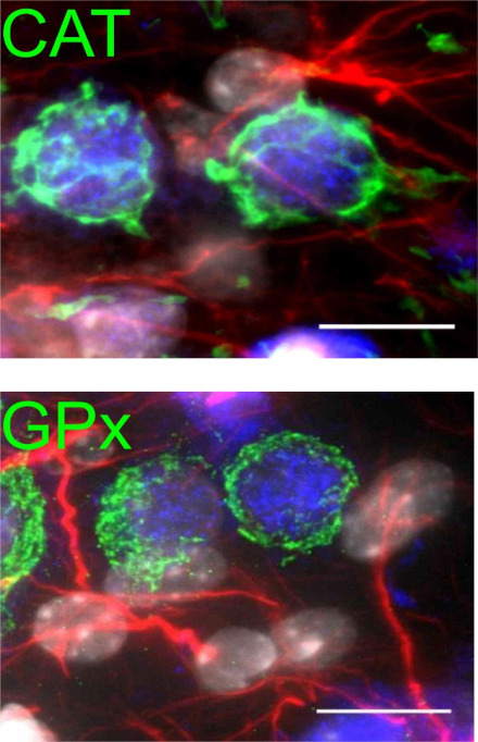
Immunohistochemistry is routinely performed to visualize potential changes in the expression of proteins. We can label up to 4 fluorophores in the same tissue to simultaneously examine multiple target proteins and their (co-)expression. Photos are then taken with conventional and laser scanning confocal microscopes. Quantification of protein expression is done via western blot analysis.
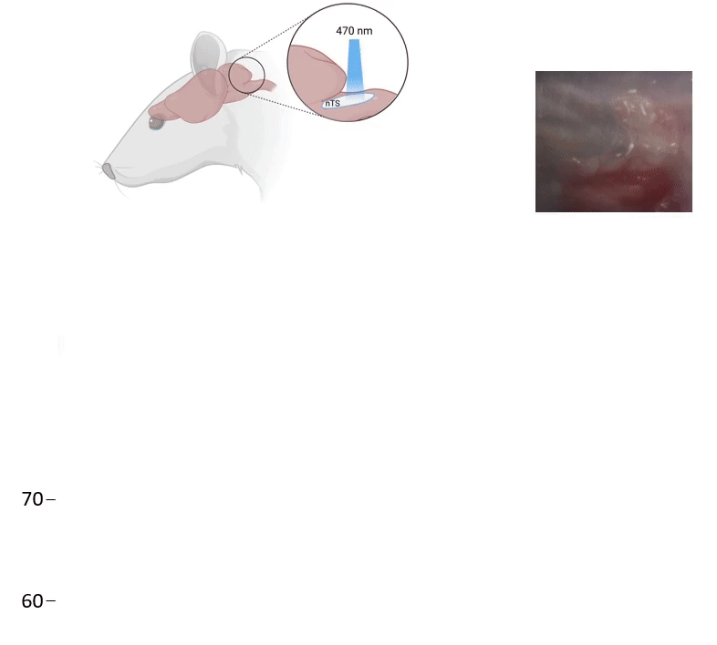
In vitro and in vivo Optogenetics: Using adeno-associated viruses, we can express channels that respond to blue light directly in respiratory regions of the nTS. We then can alter the activity within the nTS, increase breathing, and address our questions about how Alzheimer's impairs the respiratory system.